






Fusion prostate biopsy - how it works and why it is a breakthrough
A proper prostate biopsy plays a key role in the early detection of prostate cancer, which is among the most common cancers in men. While in the past mainly the classic systematic biopsy through the rectum was used, modern medicine now offers a more precise method - a fusion prostate biopsy performed in a targeted way through the perineum (the skin between the testicles and the anus). This technology significantly increases the chance of detecting a tumour at an early stage, while reducing the number of unnecessary samples, lowering the risk of infection and, not least, when a small lesion is diagnosed early we can offer the patient focal treatment of the diagnosed prostate cancer.
Main advantages of fusion biopsy
- significantly higher diagnostic accuracy
- targeted sampling from suspicious areas
- fewer needle punctures
- lower risk of missing a tumour
- suitable also after a previous negative biopsy
Fusion prostate biopsy: the most precise way to diagnose prostate cancer
Fusion biopsy is an advanced diagnostic method that combines two imaging technologies - magnetic resonance imaging (MRI) and ultrasound. MRI can image the prostate tissue very precisely and identify suspicious lesions in the prostate. Ultrasound is then used during the procedure itself to guide the needle.
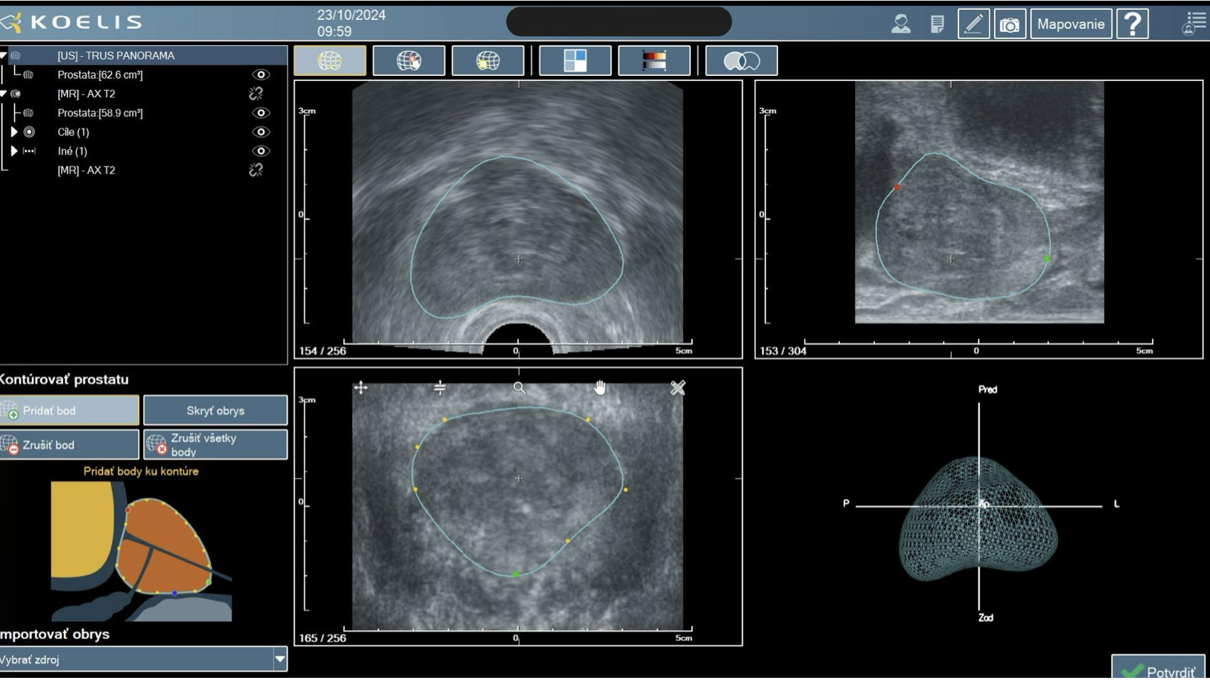
Using special software, these two images are fused, i.e. overlaid, creating a three-dimensional map of the prostate. The doctor can thus see exactly where the risk area is located and can take samples specifically from it.
How the examination works
Before the biopsy itself, the patient must undergo an MRI examination of the prostate. Based on this examination, the radiologist marks the areas that look like potential tumour lesions. These data are then uploaded to the Koelis Trinity system - a device for transperineal targeted fusion prostate biopsy.
During the biopsy itself, the patient lies on the examination table and the doctor inserts an ultrasound probe into the rectum. The computer then links the live ultrasound image with the MRI scans. The doctor can thus see exactly where to insert the biopsy needle.
The samples are taken in a very targeted way, i.e. from the specific places that the MRI marked as risk areas. The whole procedure usually takes 20 to 30 minutes and is performed under local anaesthesia.
Why fusion biopsy is even more precise with Koelis Trinity technology
In a classic biopsy, the samples are taken "blindly" from various parts of the prostate according to a standard scheme. This means the tumour may go unnoticed if the needle does not hit the right spot.
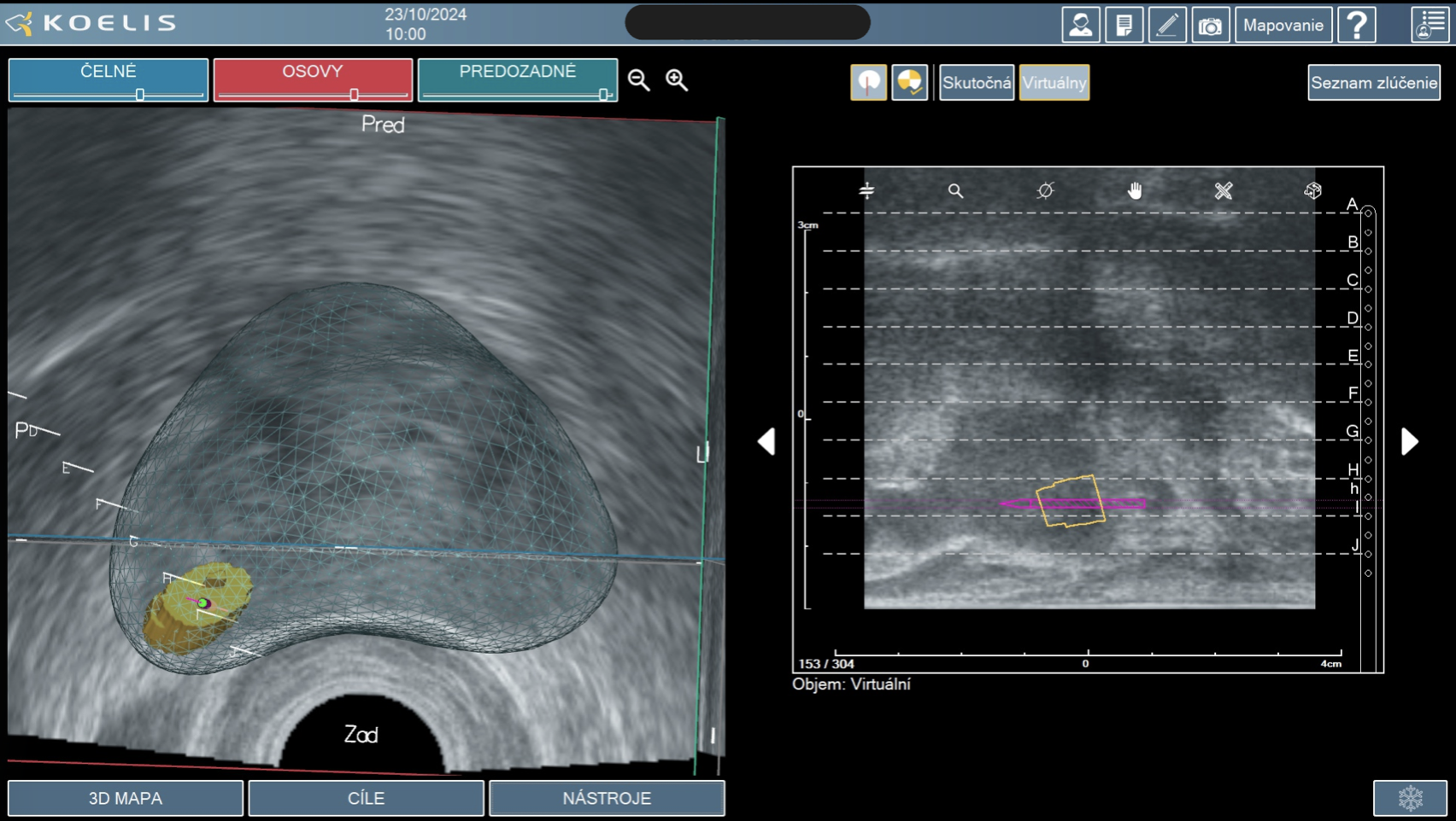
Modern clinics today use advanced fusion systems, which include the Koelis Trinity platform. This system takes the accuracy of prostate biopsy a step further, because it allows a very precise link between the MRI scans and the ultrasound image in real time.
Before the procedure itself, the patient undergoes an MRI examination of the prostate, which can identify suspicious lesions in the prostate tissue. These scans are then uploaded to the Koelis Trinity system, which links them to the live ultrasound image during the procedure. Thanks to special software, a three-dimensional map of the prostate is created, allowing the doctor to precisely locate the risk areas.
One of the great advantages of the Koelis Trinity system is also its 3D navigation and real-time tracking of the biopsy needle position. The doctor can thus see exactly from which place a sample was taken and can target specific suspicious lesions.
The technology also allows:
- very precise targeting of the biopsy at suspicious lesions from the MRI,
- a three-dimensional view of the prostate throughout the procedure,
- precise navigation of the biopsy needle in real time,
- recording of the sampling sites for future check-ups or repeat biopsies,
- a higher probability of detecting a clinically significant tumour.
Thanks to these possibilities, fusion biopsy with Koelis Trinity technology is one of the most precise diagnostic methods when prostate cancer is suspected. For the patient, this means greater certainty of a correct diagnosis, fewer unnecessary samples and better planning of further treatment.
Who is fusion prostate biopsy most suitable for
Fusion prostate biopsy is a modern diagnostic method. It is most often recommended for patients who have an increased risk of prostate cancer or in whom there is a suspicious finding on imaging examinations.
Patients with elevated PSA
- an elevated PSA level can be the first signal that a pathological process is taking place in the prostate. Fusion biopsy allows a more precise determination of whether it is a tumour disease or another problem, for example prostatitis or benign enlargement of the prostate.
Patients with a suspicious finding on MRI
- if magnetic resonance imaging shows a suspicious area in the prostate, fusion biopsy allows targeted sampling precisely from this area, which increases the probability of detecting a tumour.
Patients with a previous negative biopsy
- some patients have repeatedly elevated PSA, but a classic biopsy has not confirmed a tumour. In such cases, fusion biopsy can be significantly more precise, because it allows specific suspicious lesions to be targeted.
Patients with a higher risk of prostate cancer
Fusion biopsy is often recommended for men:
- with a genetic predisposition,
- with a family history of prostate cancer,
- with significant changes on the MRI examination,
- or with a rapid rise in the PSA value.
Thanks to the precise imaging of the prostate, the doctor can better decide on the further course of treatment.
Frequently asked questions (FAQ)
Is fusion prostate biopsy painful?
The examination is performed under local anaesthesia, which significantly reduces discomfort during the procedure. Most patients report only mild pressure or short-term discomfort.
How long does the examination itself take?
The procedure itself usually takes about 20 to 30 minutes. After a short observation the patient can go home.
Is hospitalisation necessary?
In most cases it is an outpatient procedure that does not require hospitalisation.
When will the biopsy results be known?
The tissue samples are sent for histological examination. Results are usually available within 14-21 working days.
Can I return to normal activities after the examination?
In most cases the patient can return to normal activities the very next day. However, a short period of rest and adequate fluid intake are recommended.
What side effects may occur?
After the biopsy the following may appear briefly:
- blood in the urine,
- blood in the ejaculate,
- mild burning when urinating.
These symptoms are usually temporary and resolve spontaneously.
We perform the examination at our clinic, and you can find out whether this examination is suitable for you as well here.
Latest news